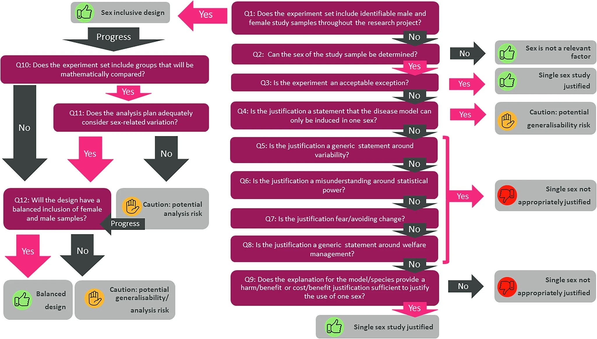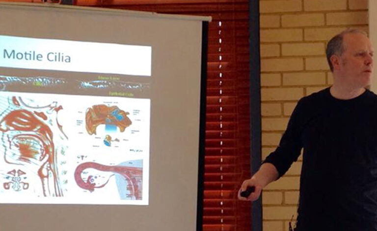
On 6 June 2015 Dominic Norris spoke to a support group for Primary Ciliary Dyskinesia, a genetic disorder of cilia, about a link between cilia and a reversed organ arrangement.
Discussing cilia research with PCD patients On 6 June 2015 Dominic Norris spoke to a support group for Primary Ciliary Dyskinesia, a genetic disorder of cilia, about a link between cilia and a reversed organ arrangement. Dominic Norris, the leader of our cilia, development and disease research group, spoke to a room full of patients and their carers on 6 June at the annual general meeting of the Primary Ciliary Dyskinesia (PCD) Family Support Group. His talk focused on the role of cilia in defining the arrangement of our internal organs, and how this process can be disrupted in PCD patients. Motile cilia are tiny, hair-like structures that protrude from many different cell types. Their roles vary, but probably the best known example is the ‘muco-ciliary escalator’ – the movement of mucus and any debris caught in it out of the lungs by the sweeping motion of the cilia that line the airways. PCD is a rare inherited disorder caused by dysfunctional motile cilia. This means patients have trouble clearing their lungs, ears and sinuses, making them prone to recurring bacterial infections. If left untreated, chest infections can lead to a type of severe lung damage known as bronchiectasis. They may also have fertility problems, and around half have ‘situs inversus’, where the internal organisation of the left and right sides of the body are the opposite way around to normal. While the human body is largely symmetrical, there are certain organs that have a preferred orientation. In those with situs inversus, it is as if a mirror was held up to the body – the heart, for example, is over to the right rather than its normal position on the left. Perhaps surprisingly, this total reversal of a person’s organ arrangement causes few problems, and accounts exist of the condition only being discovered during an X-ray, operation or post-mortem. Situs inversus is the result of cilia malfunctioning during the third week of embryonic development. A small dimple forms at one end of the embryo, known as ‘the node’, filled with cilia. These cilia normally swirl around in a circular movement, driving a leftward flow across the node, which is detected by cells around the rim. This leads to differences in gene expression on the left and right side of the embryo, resulting in the baby being born with a normal organ arrangement. In PCD patients these cilia are faulty, meaning the flow is disrupted, which leads to situs inversus 50% of the time. Dominic spoke about this link between PCD and situs inversus, and how his team investigate the genes involved in this process of left-right patterning by studying it in mice. The session went down well with the audience, with the majority saying they found it interesting, useful and informative.